
Podoplanin, a specific marker for lymphatic endothelial cells
Issuing time 2025-05-28 12:23:04
Lymphatic Vessel Invasion (LVI)
It refers to the invasion of malignant tumor cells into the lymphatic vessels and their growth in lymphatic vessels. It is one of the important pathways for local invasion and distant metastasis of tumor cells. After tumor cells invade the lymphatic vessels, they can reach local lymph nodes or more distant sites along with the flow of lymph fluid, thus lead to the spread and metastasis of the tumor.
The Process of Tumor Cells Entering Lymphatic Vessels
Tumor cells first detach from the primary tumor site. Then they go through the basement membrane and they invade the surrounding lymphatic vessels. After entering the lymphatic vessels, the tumor cells may form cancer emboli within the lumen or attach to the endothelial cells of the lymphatic vessels, and then flow with the lymph fluid to the lymph nodes or other organs.
It is difficult to distinguish lymphatic vessels and blood vessels through HE staining
There are subtle structural differences between blood vessels and lymphatic vessels. In HE staining, blood vessels and lymphatic vessels are distinguished by observing whether there are red blood cells in the lumen of the blood vessels. Sometimes, due to the slicing direction or vasoconstriction, red blood cells may not be visible in the blood vessels. Occasionally, red blood cells may infiltrate into the lymphatic vessels due to inflammation or injury.
Structure and Function of Podoplanin (PDPN)
Podoplanin (PDPN) belongs to the mucin-type transmembrane glycoprotein family, with a molecular weight of approximately 40 kD. It has a unique structure, consisting of a relatively long extracellular domain, a transmembrane domain, and a shorter intracellular domain (Figure 1). As a cell surface receptor, Podoplanin is involved in the generation and maintenance of lymphatic vessels, as well as the process of tumor cell metastasis through the lymphatic vessels.
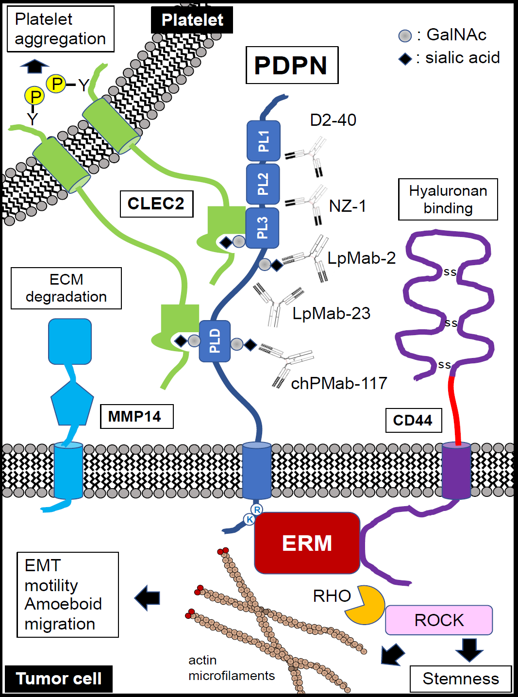
Figure 1. Schematic representation of podoplanin (PDPN) structure and functions.
PDPN is a type I transmembrane glycoprotein consisting of an extracellular domain, a transmembrane portion, and a short cytoplasmic tail. The PDPN extracellular domain contains PLAG1-3 (PL1, PL2, and PL3) domains and PLAG-like domain (PLD). C-type lectin-like receptor 2 (CLEC-2), a platelet receptor, recognizes both the sialylated PLAG3 domain and PLD with the adjacent PDPN peptides, inducing CLEC-2 tyrosine phosphorylation and platelet aggregation. The intracellular domain of PDPN contains basic residues (RK), which function as binding sites for ezrin, radixin, and moesin (ERM) family proteins that modulate RHO GTPase activity and promote actin cytoskeleton reorganization to promote cell migration, motility, and EMT. PDPN interacts with hyaluronan receptor CD44 and matrix metalloproteinase 14 (MMP14), promoting hyaluronan-binding and extracellular matrix (ECM) degradation, respectively. Anti-PDPN mAb NZ-1 recognizes the PLAG2/3 domain, exhibits a neutralizing activity for PDPN–CLEC-2 interaction, and inhibits PDPN-induced platelet aggregation and metastasis. Anti-PDPN mAb D2-40 identifies the PLAG1/2 domain and is widely employed for immunohistochemistry. A cancer-specific mAb (CasMab) to PDPN, LpMab-2, recognizes a glycopeptide (Thr55-Leu64) of human PDPN. A CasMab to PDPN, LpMab-23 recognizes a naked peptide of human PDPN (Gly54–Leu64), especially Gly54, Thr55, Ser56, Glu57, Asp58, Arg59, Tyr60, and Leu64 of PDPN, and is a critical epitope of LpMab-23. A CasMab to PDPN, chPMab-117 recognizes the glycopeptide of PDPN (Ile78-Thr85), which includes O-glycosylated Thr85.
Source: Suzuki H, Kaneko MK, Kato Y. Roles of podoplanin in malignant progression of tumor. Cells. 2022 Feb 7;11(3):575.
https://doi.org/10.3390/cells11030575
D2 – 40 is a monoclonal antibody of podoplanin
Podoplanin is a specific marker for lymphatic endothelial cells, and D2-40 is a monoclonal antibody of podoplanin. By detecting podoplanin through immunohistochemical staining, the distribution and morphology of lymphatic vessels can be clearly observed. This can help doctors assess the risk of tumor metastasis and provide a crucial basis for formulating clinical treatment plans. In addition, detecting Podoplanin using the D2-40 antibody can also distinguish lymphangiomas from other soft tissue tumors.
We, Dartmon, provide this D2-40 to assist with your diagnosis.

Intense staining of the follicular dendritic cells (red arrows) in the germinal center can be seen. Basal squamous epithelial cells (black arrows) has been strongly stained.

The endothelial cells lining the lymphatic vessels (red arrow) show intense and distinct staining, while the vascular endothelium (black arrow) shows no staining. In the muscular layer, the Cajal cells (green arrow) show moderately positive staining.
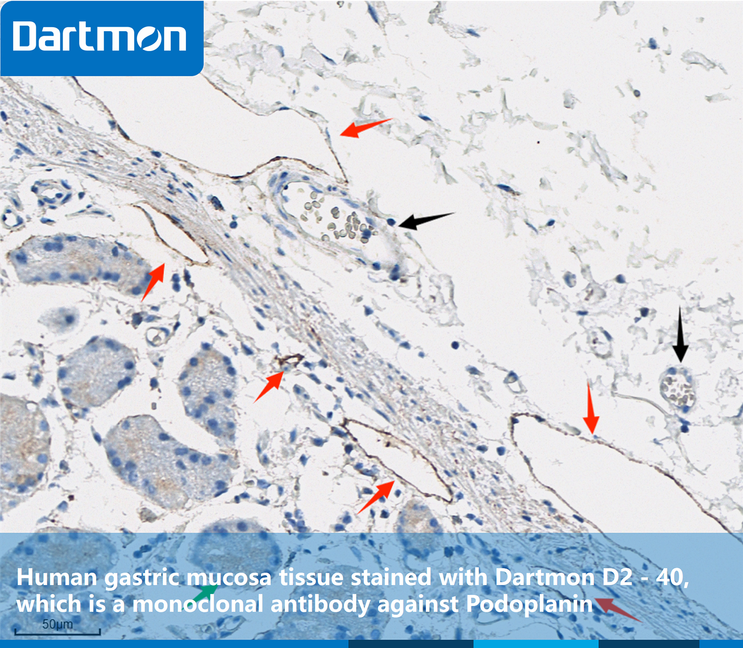
The endothelial cells lining the lymphatic vessels (red arrow) show intense and distinct staining, while the vascular endothelium (black arrow) shows no staining. Due to the mucus secretion of the glands, there is a slightly acceptable background staining (green arrow).

The lymphatic endothelial cells (red arrow) show strong cytoplasmic expression, and the vascular endothelium shows no staining (black arrow); In the myoepithelial cells related to benign ducts and lobules (green arrow), podoplanin is expressed in a weak patchy cytoplasmic pattern.

Colorectal tumor cells (black arrow) show negative expression, while fibroblasts (red arrow) dispersed in the stroma or forming a reticular network (reticular pattern) show strong positive expression.
Product infomation:
Anti-Podoplanin (DA051)
Cat. No. : MMB1A092
Usage pattern: Manual or device utilization
Ready-to-use: 3ml, 6ml, 10ml
Concentrated: 0.1ml, 0.5 ml, 1ml

