Pax8: A Commonly Used Renal Marker and Latest Research Advances
Issuing time 2025-07-10 15:59:32
PAX8 is a member of the mammalian Paired-box family of genes that includes 9 (PAX1–9) different genes and plays a crucial role in the organogenesis of the kidney, thyroid gland, and Müllerian organs [1–4].

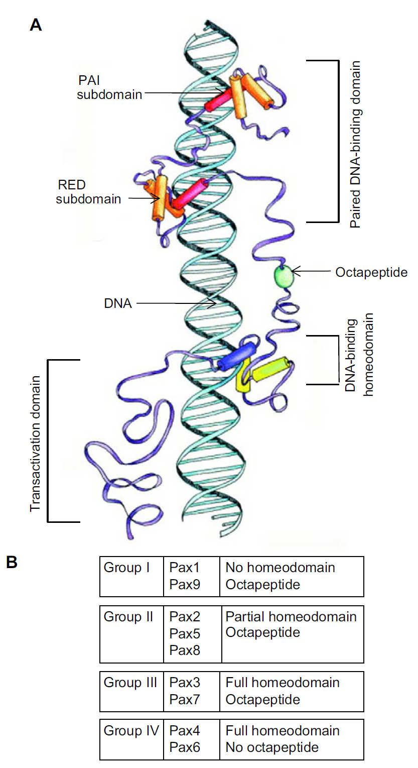
Fig. 1 Paired-box (PAX) protein structure [5]
PAX proteins are characterized by their conserved, paired DNA-binding domain, which is composed of the RED and PAI subdomains. b The partial or full inclusion of a DNA-binding homeodomain and/or octapeptide linker subdivides PAX proteins into Group 1, Group 2, Group 3, or Group 4 [5].
Among various embryonic genes expressed during kidney development, the fundamental role in branching morphogenesis and nephron differentiation belongs to PAX8 transcription factor [6].
Using multiomic analysis of human renal organoids and fetal kidneys, Ng-Blichfeldt et al. map transcriptional dynamics controlling the emergence of nephron epithelia from mesenchymal progenitors during human kidney development, revealing critical roles for PAX8 and transient Wnt signaling [7].

Fig. 2, PAX8 initiates renal MET downstream of Wnt/b-catenin signaling [7] MET: mesenchymal-to-epithelial transition
In diagnostic pathology, PAX8 immunohistochemistry (IHC)—in combination with other markers—is often used to determine the origin of tumors that are difficult to classify by morphology alone. Detectable PAX8 expression is considered a strong argument for a tumor origin from the kidney, thyroid, or inner female genital tract [8, 9]

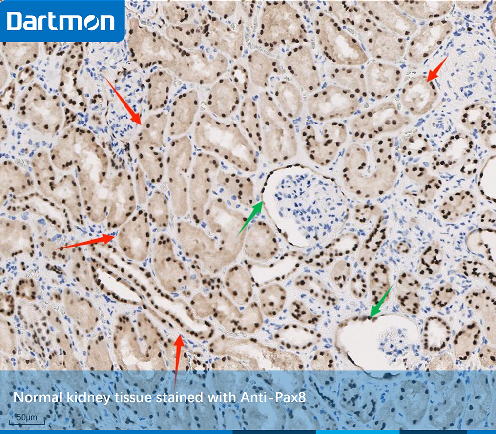
Fig. 3, In the kidney, strong nuclear PAX8 staining of cells can be seen in proximal and distal tubuli, collecting ducts (Red Arrow) and epithelial cells of the parietal membrane of the Bowman’s capsule (Green Arrow).

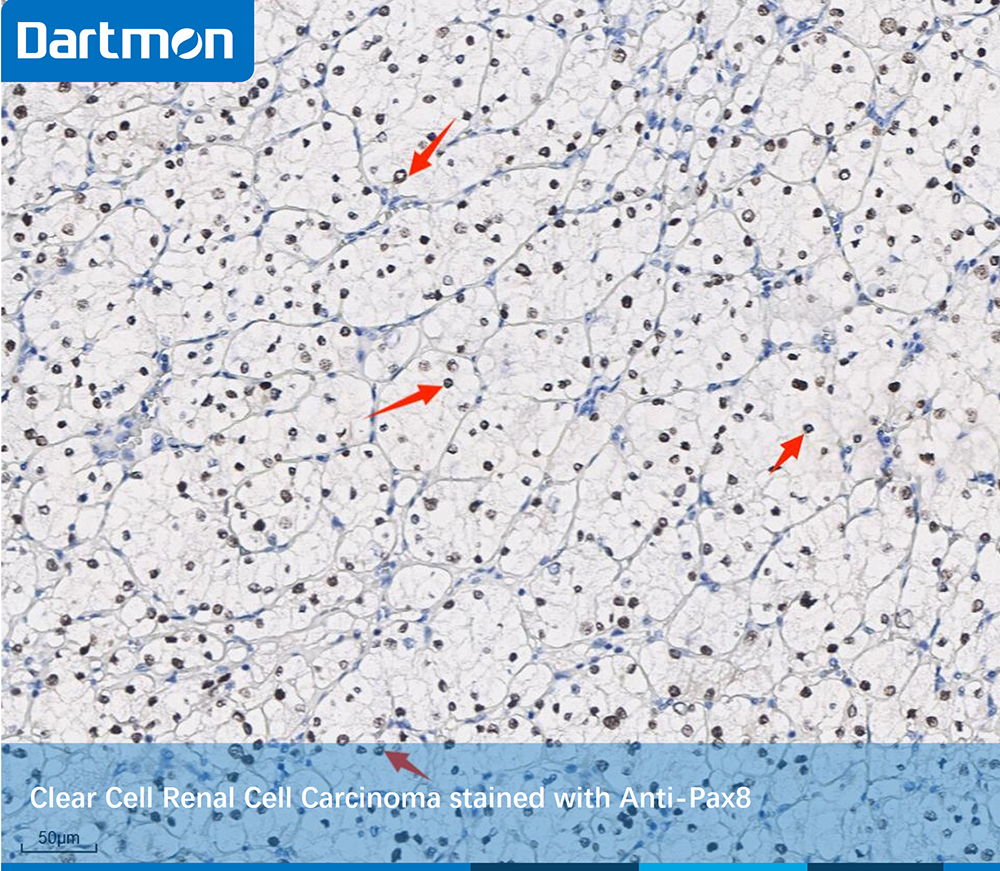
Fig. 4, PAX8 immunostaining in Clear Cell Renal Cell Carcinoma(ccRCC). Almost all tumor cells (red arrows) showed moderate to strong nuclear staining reactions.

We, Dartmon, provide you the best-in-class Pax8 antibody!
Product name: Anti- Pax8 (DA099)
Cat. No. : RMB1A110
Usage pattern: Manual or device utilization
Ready-to-use: 3ml, 6ml, 10ml
Concentrated: 0.1ml, 0.5 ml, 1ml
References
1. Lang, D.; Powell, S.K.; Plummer, R.S.; Young, K.P.; Ruggeri, B.A. PAX genes: Roles in development, pathophysiology, and cancer. Biochem. Pharmacol. 2007, 73, 1–14.
2. Mansouri, A.; Hallonet, M.; Gruss, P. Pax genes and their roles in cell differentiation and development. Curr. Opin. Cell Biol. 1996, 8, 851–857.
3. Plachov, D.; Chowdhury, K.; Walther, C.; Simon, D.; Guenet, J.L.; Gruss, P. Pax8, a murine paired box gene expressed in the developing excretory system and thyroid gland. Development 1990, 110, 643–651.
4. Grimley, E.; Dressler, G.R. Are Pax proteins potential therapeutic targets in kidney disease and cancer? Kidney Int. 2018, 94, 259–267.
5. Blake JA, Ziman MR (2014) Pax genes: regulators of lineage specification and progenitor cell maintenance. Development 141:737.
6. Narlis, M.; Grote, D.; Gaitan, Y.; Boualia, S.K.; Bouchard, M. Pax2 and Pax8 regulate branching morphogenesis and nephron differentiation in the developing kidney. J. Am. Soc. Nephrol. 2007, 18, 1121–1129.
7. Ng-Blichfeldt, J. P., Stewart, B. J., Clatworthy, M. R., Williams, J. M., & Röper, K. (2024). Identification of a core transcriptional program driving the human renal mesenchymal-to-epithelial transition. Developmental cell, 59(5), 595-612.
8. Ozcan A, Shen SS, Hamilton C, Anjana K, Cofey D, Krishnan B, Truong LD (2011) PAX 8 expression in non-neoplastic tissues, primary tumors, and metastatic tumors: a comprehensive immunohistochemical study. Mod Pathol 24:751–764. https://doi.org/10.1038/modpathol.2011.3
9. Tacha D, Zhou D, Cheng L (2011) Expression of PAX8 in normal and neoplastic tissues: a comprehensive immunohistochemical study. Appl Immunohistochem Mol Morphol 19:293–299. https://doi.org/10.1097/PAI.0b013e3182025f66


